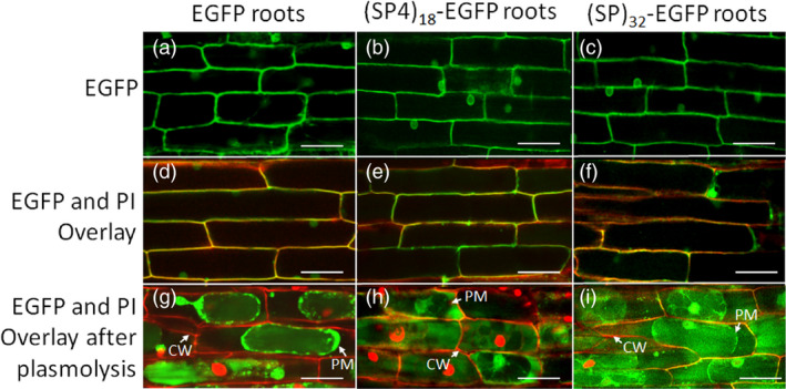
Figure 3.

Fluorescence micrographs of hairy roots expressing EGFP, (SP4)18‐EGFP and (SP)32‐EGFP. The hairy roots grown in petri dish for 12 days were inspected using a laser‐scanning confocal microscope with a 40× water‐immersion objective. The cell wall of the root tissues was stained with propidium iodide (PI). (a–c) Hairy root images detected under green fluorescence channel (488 nm excitation with 525/50 nm filter); (d–f) Overlaid images captured under both green fluorescence channel and red fluorescence channel (543 nm excitation with 595/50 nm filter); (g–i) Hairy root cells plasmolysed with 800 mm mannitol. The images were then detected under both green fluorescence and red fluorescence channels and overlaid. CW, cell wall; PM, plasma membrane. Scale bar = 50 μm.
