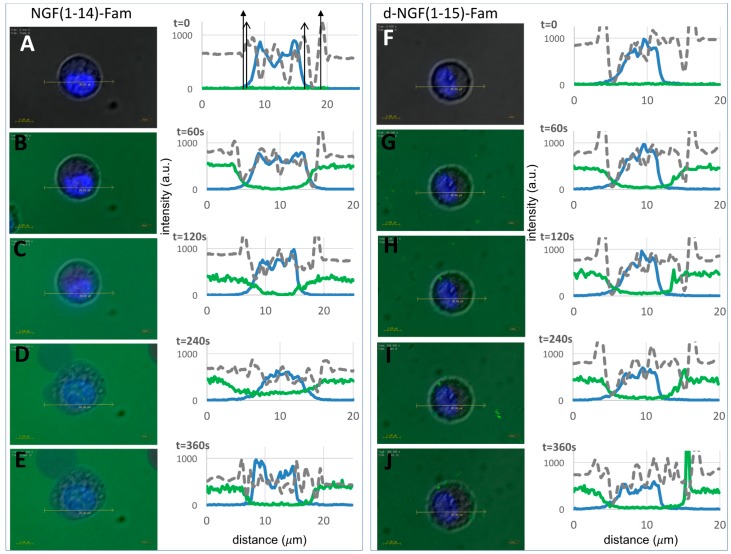
Figure 11.
Live cell imaging by confocal microscopy of Fam-labeled NGF(1-14) and d-NGF(1-15) internalization. Merged optical bright field (grey) and confocal (blue: nuclear staining, λex/em = 405/425–475 nm; green: Fam, λex/em = 488/500–530 nm) micrographs and corresponding section analysis for PC12 before (A,F) and after (60 s, 120 s, 240 s, 360 s) the addition of 10 μM NGF(1-14)-Fam (B–E) or 10 μM d-NGF(1-15)-Fam (G–J), respectively. The solid and open arrows in the top left plot guide the eye for the cell and nuclear membranes, respectively.

