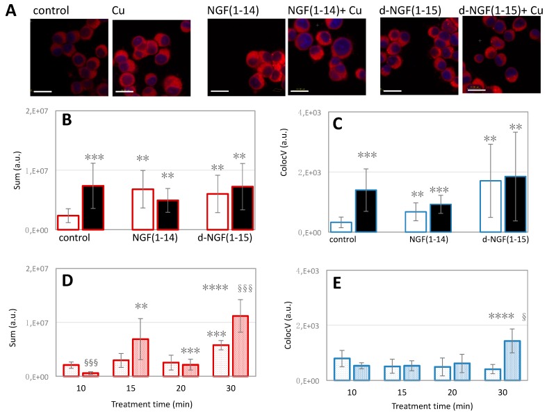
Figure 12.
The effect of d-NGF(1-15) and NGF(1-14) peptides on copper homeostasis. In (A): representative merged confocal microscopy images of coppersensor-1 (red) and Hoechst33342 ( blue) stained cells after 15 min of treatment with 10 μM NGF(1-14) or d-NGF(1-15), either in the absence or in the presence of 1 µM CuSO4. Untreated and 1 µM CuSO4 treated-cells are included as negative and positive controls, respectively (scale bar = 10 μm). (B,C): Quantitative analysis of the confocal micrographs for total cytoplasmic Cu+ (B) and nuclear Cu+ (C) in the cells incubated with the peptides in basal medium ( ,
,  ) or copper-supplemented medium (
 ,
,  ). In (D,E): quantitative analyses of total intracellular copper (D) or nuclear-confined copper (E) after PC12 cells incubation for 10, 15, 20 or 30 min with 10 μM NGF(1-14) (cytoplasm:
  ; nuclei:
  ) or 10 μM d-NGF(1-14) (cytoplasm:
  ; nuclei:
  ). ** p < 0.01, *** p < 0.001, versus control.; ** p < 0.01, *** p < 0.001, **** p < 0.0001 versus the preceding incubation time; § p < 0.05, §§§ p < 0.001 versus [NGF(1-14)].

