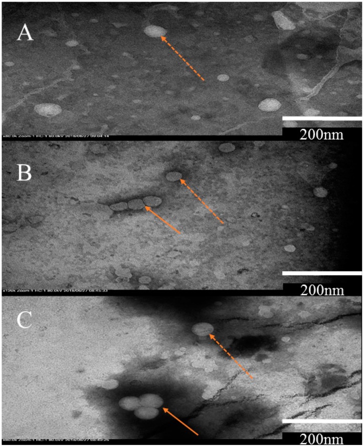
Figure 2.
Transmission electron microscopy (TEM) analysis of liposomes prepared by; (A) homogenization method, (B) Ultrasonication and (C) Mozafari method. Sample of unilamellar vesicles are shown with a dotted arrow while aggregates are indicated by solid arrows. Scale bar in the figure A, B and C indicates 200nm, which represents the size of vesicle.

