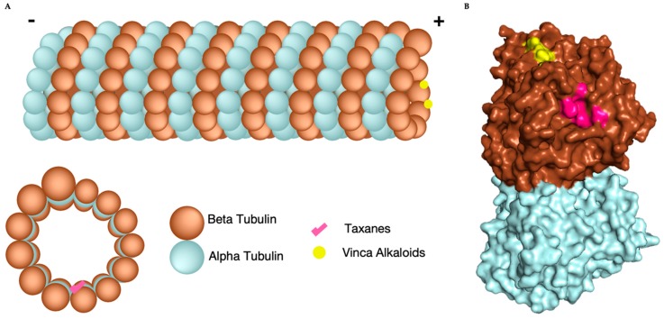
Figure 1.
Microtubule composition, structure, and microtubule targeting agents (MTA) binding sites. (A) Cartoon of microtubule consisting of 13 protofilaments formed from alpha (blue) and beta (brown) tubulin heterodimers. Vinca alkaloids (yellow) bind to the + end of the microtubule and taxanes (pink) bind inside the lumen (lower left). (B) Space Filling model an of alpha (Blue) beta (Brown) heterodimer with vinca alkaloid binding site (yellow) and taxane binding site (pink) located on beta tubulin [PDB: 3J8Y].

