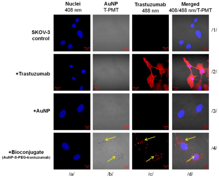
Figure 4.
Internalization of free AuNPs and AuNP-S-PEG-trastuzumab bioconjugate by SKOV-3 cells determined by confocal microscopy. SKOV-3 cells that were untreated or treated only with trastuzumab served as positive and negative controls, respectively. Fluorescence signals indicate: (red)—subcellular trastuzumab distribution; (blue)—nuclei intracellular localization. Au-containing particles (dark spots) were visualized with a transmitted light detector (T-PMT). Merged images are presented in column d. Arrows mark the subcellular localization of the AuNP-S-PEG-trastuzumab bioconjugate.

