Figure 4.
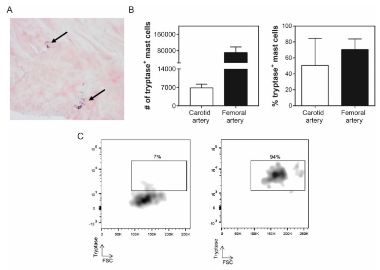
Tryptase content of mast cells in human atherosclerotic plaques. (A) Immunohistochemical tryptase staining of a human atherosclerotic plaque. Arrows indicate activated mast cells. (B) Number (left panel) and percentage (right panel) of mast cells containing tryptase in human carotid and femoral arteries based on flow cytometry. (C) Flow cytometry plot examples of tryptase+ mast cells, that is, left panel: low tryptase expression (7% of the mast cell population) vs. right panel: high tryptase expression (94% of the mast cell population). Values are depicted as mean ± SEM.

