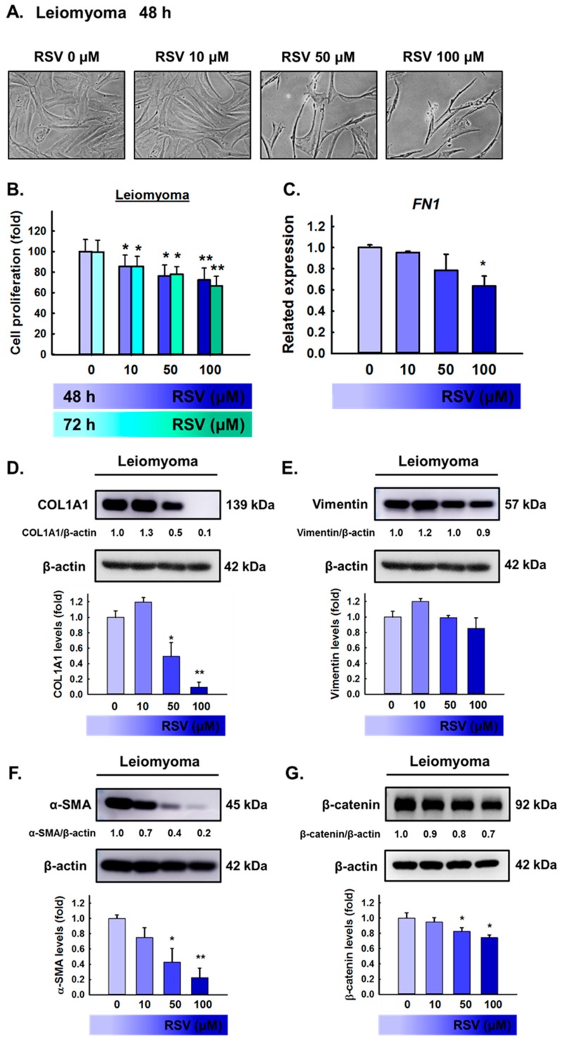
Figure 4.
Cytotoxic effects of resveratrol (RSV) on primary human leiomyoma cells. Leiomyoma cells were exposed to either vehicle (dimethyl sulfoxide; DMSO) or RSV (10–100 μM) for 48 h or 72 h. (A) Morphology of leiomyoma cells after the indicated treatment (magnification, ×200). (B) Cell proliferation was measured using a 3-(4,5-dimethylthiazol-2-yl)-2,5-diphenyltetrazolium bromide (MTT) assay. (C) RNA samples were isolated from leiomyoma cells treated with RSV (0–100 μM) and subjected to quantitative reverse transcription–polymerase chain reaction (qRT–PCR) using primers specific for fibronectin (FN1). (D–G) Leiomyoma cell lysates were separated using sodium dodecyl sulfate polyacrylamide gel electrophoresis (SDS–PAGE) and analyzed using Western blot with anti-COL1A1, vimentin, α-SMA, and β-catenin. β-actin was used as a loading control. The values of the band intensity represent the densitometric estimation of each band normalized to β-actin. Protein quantification of COL1A1, vimentin, α-SMA, and β-catenin expression in leiomyoma cells is shown in the bar graph. The results are expressed as the means ± SD of three independent experiments; * p < 0.05, ** p < 0.001, as compared with the control.

