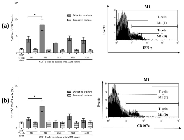
Figure 6.
Increased IFN-γ and CD107a expression by CD8+ T-cells following co-culture with macrophage subsets is contact-dependent. Macrophage subsets were co-cultured either directly with autologous CD8+ T-cells or were separated by a transmembrane. After 24 h of culture, intracellular expression of IFN-γ was evaluated in CD8+ T-cells by flow cytometry. (a) The proportion (%) of IFN-γ+ CD8+ T-cells following either direct co-culture (grey bars) or co-cultures separating MDM from CD8+ T-cells in a transwell (hatched bars) is shown in the summary graph (n = 3). A representative histogram depicts intracellular IFN-γ expression by CD8+ T-cells in direct and transwell co-culture with M1 macrophages and includes a trace for CD8+ T-cells cultured alone (filled black area). (b) After 48 h of direct or indirect co-culture of macrophage subsets with CD8+ T-cells, the expression of CD107a was evaluated by flow cytometry. A representative histogram compares the CD107a expression of CD8+ T-cells in direct and transwell co-culture with M1 macrophages and includes a trace for CD8+ T-cells cultured alone (filled black area). Statistical significance was determined by a Student’s t-test (p ≤ 0.05), and significant p-values are indicated with an asterisk “*”.

