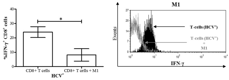
Figure 7.
M1 macrophages derived from HCV-infected individuals with minimal liver fibrosis decrease the proportion of IFN-γ+ CD8+ T-cells in co-culture. After differentiation from MDMs derived from blood monocytes from HCV-infected individuals, M1 macrophages were co-cultured for 24 h with autologous CD8+ T-cells isolated from frozen peripheral blood mononuclear cells. Intracellular IFN-γ expression in CD8+ T-cells was then evaluated by flow cytometry. The proportion of IFN-γ+ CD8+ T-cells in co-cultures was compared to that of CD8+ T-cells cultured alone (n = 7). Representative histograms of IFN-γ expression by CD8+ T-cells co-cultured with M1 macrophages (unfilled area) superimposed over CD8+ T-cells cultured alone (black filled area) are also shown. Statistical significance was determined by a Student’s t-test (p ≤ 0.05), and statistical significance is indicated with an asterisk “*”.

