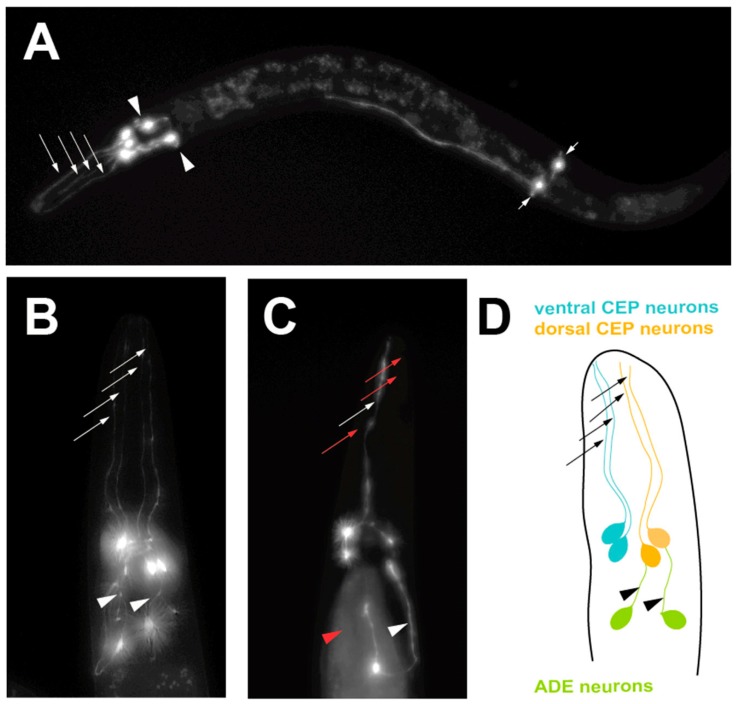
Figure 2.
A progressive, age-dependent C. elegans α-syn model of PD [18]. (A) A worm expressing solely Green Fluorescent Protein (GFP) in the 8 DA neurons, with GFP being driven under the dopamine neuron-specific dat-1 promoter for visualization. Arrows with long tails indicate the 4 CEP neuron processes, arrowheads indicate the 2 ADE neuron processes and arrows with short tails indicate the 2 PDE neuron processes. (B) A worm expressing both GFP and human, wild-type α-syn in the DA neurons being driven under the dopamine neuron-specific dat-1 promoter, allowing visualization of the 6 DA neurons located in the head region. Arrows indicate non-degenerated CEP neuron processes and arrowheads indicate non-degenerated ADE neuron processes. (C) A worm expressing both GFP and human, wild-type α-syn in the DA neurons being driven under the dat-1 promoter, allowing visualization of the 6 DA neurons located in the head region. Red arrows indicate degenerated CEP neuron processes and the red arrowhead represents a degenerated ADE neuron process. (D) A representation of the neuroanatomy of the 6 DA neurons in the head region of C. elegans. Ventral and dorsal CEP neurons are shown, along with the ADE neurons.

