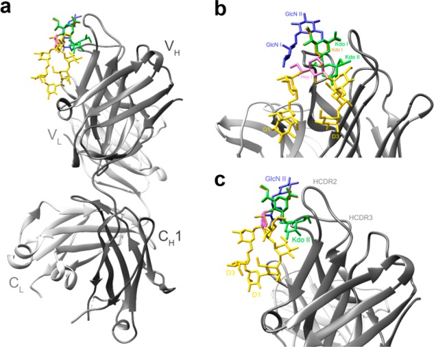
Figure 3.

Interaction of HIV-broadly neutralizing antibody PGT128 with a model of the undecasaccharide. (a) Overview of the modeled interaction. The crystal structure complex of PGT128 with heptamannoside NIT68A13,34 was used as template to create the model. The heavy and light chains of the PGT128 Fab are shown in a ribbon representation in dark gray and light gray, respectively. The modeled undecasaccharide is shown in stick representation, with the NIT68A heptamannoside in yellow, Kdo disaccharide in green, and the GlcN disaccharide in blueish purple. (b) Close-up view showing the modeled interaction of PGT128 with the undecasaccharide. The constituents of the undecasaccharide are labeled. To model the Kdo disaccharide, a crystallized Hep-Kdo2 fragment35 was modeled into the PGT128 binding site by superposing the heptose residue (pink) onto the central branching mannose of the NIT68A structure. To model the lipid A disaccharide, a crystallized Kdo-GlcN2 fragment36 was superposed onto the structure using the main-chain Kdo residue (Kdo I; orange) as a guide. (c) Left side view of panel b, highlighting the assumed proximity of the side chain Kdo (Kdo II) to the antibody.
