FIGURE 2.
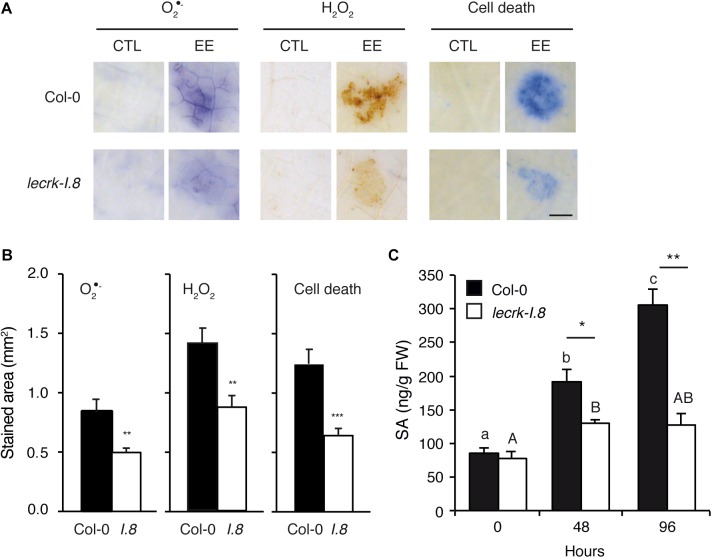
LecRK-I.8 is involved in signaling of Arabidopsis response to EE. (A) Leaves from Col-0 and leckrk-I.8 were treated with P. brassicae EE for 72 h. Histochemical staining of leaves with nitroblue tetrazolium (NBT) to detect O2∙-, 3,3-diaminobenzidine (DAB) to detect H2O2, and trypan blue to detect cell death was performed. Untreated plants were used as controls (CTL). Panels are close-up images of the spotted area. Representative photographs from several replicates are shown. Bar = 1 mm. (B) Quantification of ROS and cell death accumulation in response to EE treatment as in (A). Stained area was measured on images with ImageJ software (n = 10). Means ± SE are shown. Significant differences are indicated (Student’s t-test, ∗∗P < 0.01). I.8, lecrk-I.8. (C) Free salicylic acid (SA) was quantified in leaf discs of 10 mm diameter (n = 15) during 96 h after application of P. brassicae EE in Col-0 (black bars) and lecrk-I.8 (white bars). Means ± SE of three independent biological replicates are shown. Different letters indicate significant differences (ANOVA followed by Tukey’s honest significant difference test, P < 0.05). Significant difference between wild-type and mutant are indicated (Student’s t-test, ∗P < 0.05, ∗∗P < 0.01, ∗∗∗P < 0.001).

