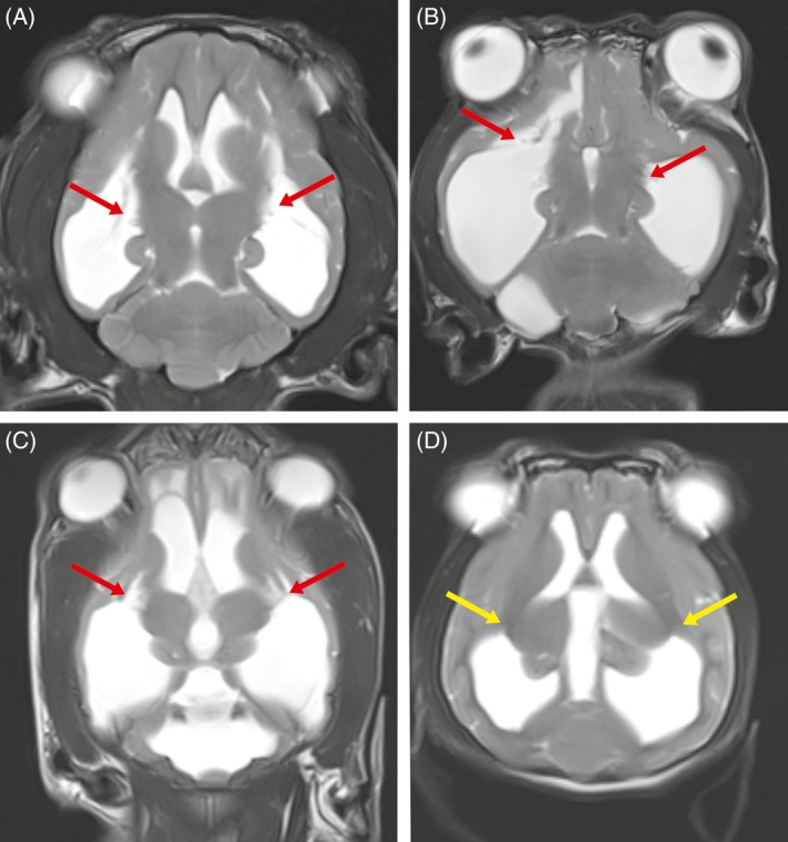
Figure 2.

Dorsal T2‐weighted magnetic resonance images through the brain of dogs with internal hydrocephalus (A: dog 4; B dog 13; C dog 40; D: dog 2). In the dogs A‐C in which blindness was diagnosed clinically, a detachment of the white matter containing the optic radiation from the thalamus was found (red arrows). The dog in image D is a dog with internal hydrocephalus but with unimpaired visual function. The yellow arrow highlights the area of the optic radiation in the internal capsule
