
Figure 2.
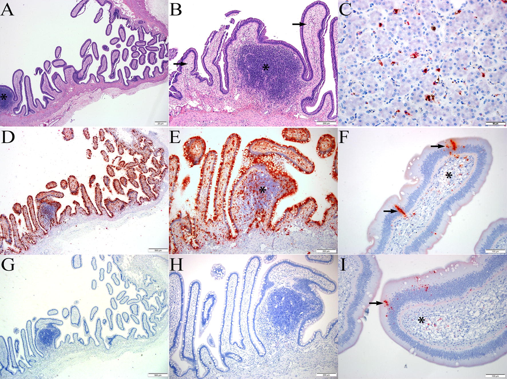
BhPyCV infection. A, B Longitudinal section of small intestine from index BhP at low (×4, A) and higher magnification (×10, B) showing a mucosal lymphoid aggregate (asterisk) and villi mildly expanded by edema and lymphocytes (arrows), H&E. D, E Serial sections from A–B showing abundant, deep red, punctate to coalescing ISH signal lining the mucosal epithelium, filling intestinal villi, and scattered in lymphoid follicle (asterisk). Hematoxylin counterstain, ×4 (D); ×10 (E). G, H Serial histologic sections from A–B, D–E. Negative control using bacterial DapB gene probe with no positive ISH signal, hematoxylin counterstain, ×4 (G); ×10 (H). C BhPyCV ISH probe on liver from index BhP showing discrete, punctate red staining within sinusoidal macrophages. Hematoxylin counterstain, ×40. F, I Medium–high magnification of intestinal villi from second BhP showing similar yet less dense and frequent staining within the epithelium (arrows) and scattered in the lamina propria (asterisk). Hematoxylin counterstain, ×20.