
Fig. 2.
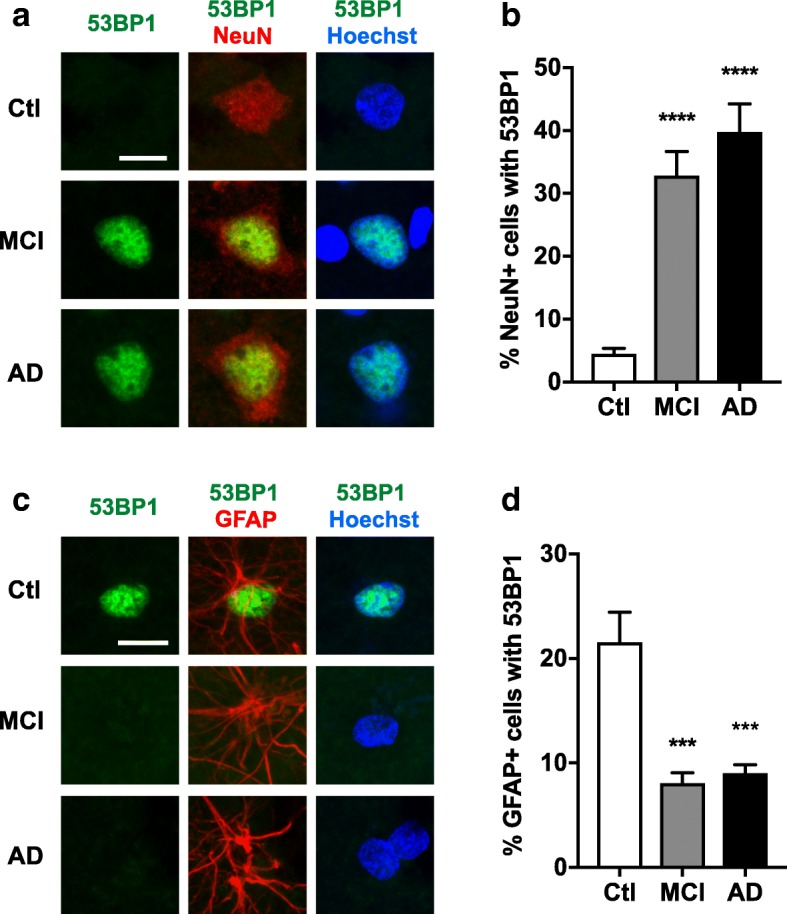
Pan-nuclear 53BP1 staining is increased in neurons and decreased in astrocytes in frontal cortex of MCI and AD cases. Frontal cortex sections from cognitively unimpaired controls (Ctl) and from MCI and AD cases were double-labeled for 53BP1 (green) and for NeuN (red) (a, b) or the astroglial marker GFAP (red) (c, d). Nuclei were stained with Hoechst 33342 (blue). a Representative confocal images of neuronal 53BP1 staining. b Proportion (%) of neurons with pan-nuclear 53BP1 staining. c Representative confocal images of astroglial 53BP1 staining. d Proportion (%) of astrocytes with pan-nuclear 53BP1 staining. n = 8 Ctl, 7 MCI, and 8 AD cases from Cohort 2. ***p < 0.001, ****p < 0.0001 vs. Ctl by one-way ANOVA and Holm-Sidak test. Scale bars: 10 μm. Bar graphs represent means ± SEM