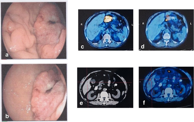
Fig. 2.
Endoscopy and PET/CT revealed GCLM. a. posterior wall of the antrum: worm-like erosion edges, visible bleeding from blood vessels and blood clots. b. small curvature of the antrum, white protrusions. c The lesion was located in the gastric antrum(SUVmax13.3), indicating GC. d Lymph node metastasis was observed below the gastric antrum (SUVmax3.3). d. Computed Tomography Image revealed a single liver metastasis, located in segments 6. e PET/CT identified localized radionuclide concentration (SUVmax3.5)

