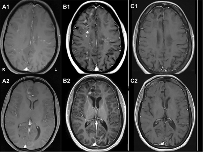
Fig. 2.
Neuroradiological Images. T1-weighted, Gadolinium enhanced images with the following findings: (a1, a2) Brain MRI at onset of neurologic symptoms shows patchy diffuse enhancement throughout both cerebral hemispheres and corpus callosum that involves cortex, juxto- and sub-cortical white matter. (b1, b2) Follow up brain MRI at the time of brain biopsy (6.5 years after onset) shows steady progression of disease with new areas of enhancement in frontal and occipital lobes as well as global cerebral volume loss (enlargement of ventricles, thinning of corpus callosum). Appearance of cavitary changes (T1 hypointensities, white arrow) suggests a more severe degree of brain injury. (c1, c2) MRI of the brain done after 7 months of tocilizumab therapy shows a remarkable decrease in the extent and number of previously enhancing lesions and no new enhancing lesions; widening of cortical sulci and ventricles is evident on follow up MRI

