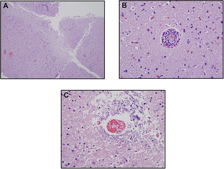
Fig. 3.

Neuropathological Findings with Hematoxylin and Eosin Staining. a The biopsy of the brain shows discrete areas of cortical necrosis. b Higher power magnification demonstrates a lymphocytic vasculopathy c pauci-inflammatory vascular thrombosis attributable to immune based endothelial cell injury. (a. Hematoxylin-eosin (H&E), 2x; b. H&E, 40x; c. H&E, 40x)
