
Figure 3.
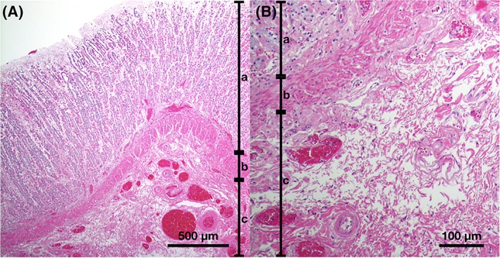
Histopathological appearance of the thickened gastric wall of a dog with low magnification image (A) and close‐up image of submucosal layer (B). There is a diffuse extensive edema in the submucosal layer separating collagen fiber. Hematoxylin and eosin. a, mucosa; b, muscularis mucosa; c, submucosa