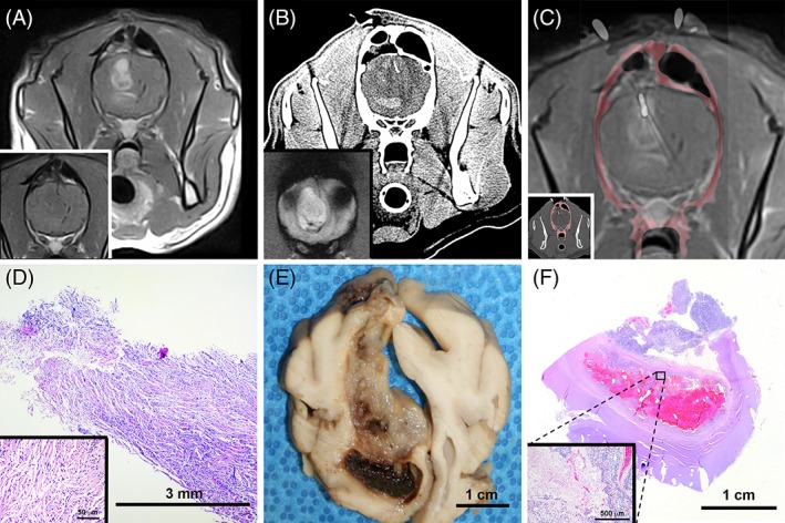
Figure 1.

Computed tomography (CT)‐guided stereotactic brain biopsy (SBB) of canine astrocytoma illustrating underestimation of tumor grade by SBB. A, Pre‐ (inset) and post‐contrast T1‐weighted (T1W) magnetic resonance image (MRI) of heterogeneously ring‐enhancing intra‐axial mass in right frontal lobe. B, Preoperative CT with hyperattenuating region of intralesional hemorrhage corresponding with hypointensity on the T2* gradient image (inset) that is avoided during biopsy. C, Coregistered post‐contrast T1W MRI and intraoperative CT scan with biopsy needle in situ (inset) within dorsal contrast‐enhancing portion of the mass. D, Core specimen from SBB depicted in (C), demonstrating features of a low‐grade fibrillary astrocytoma. The neoplasm is composed of spindloid cells with indistinct cell borders, fibrillar eosinophilic cytoplasm, and cytoplasmic processes arranged in streams and whorls (inset) around small caliber blood vessels, hematoxylin and eosin (H&E) stain. Phenotypic heterogeneity of the tumor is evident in gross (E) and subgross (F) necropsy specimens. In the ventral hemorrhagic region, features of high‐grade astrocytoma such as geographic necrosis, cellular palisading, and microvascular proliferation are apparent (F, inset; H&E stain)
