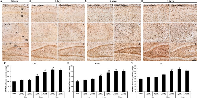
Figure 5.
Immunohistochemistry (brown color) for GFAP in the hippocampal formation of gerbils at different time after 5- and 15-min BCCAO.
(A) Sham operation; (B) 1 day after BCCAO, (C) 2 days after BCCAO, (D) 5 days after BCCAO. One day after BCCAO, the distribution of GFPA-ir astrocytes is not significantly altered in all subregions. At 2 days after BCCAO, GFAP-ir cells are significantly hypertrophied in all subregions. Five days after BCCAO, GFAP-ir cells are significantly hypertrophied in all subregions of gerbils from the 5- and 10-min BCCAO groups. Scale bar: 100 µm. (E–G) ROD of GFAP-ir cells in the CA1 (E), CA2/3 (F) and DG (G) (n = 7 at each time after BCCAO; *P < 0.05, vs. the sham operated group, #P < 0.05, vs. the prior time point of corresponding BCCAO group, †P < 0.05, vs. the same time point of 5 min-BCCAO group). The bars indicate the means ± SEM. GFAP: Glial fibrillary acidic protein; ir: immunoreactive; BCCAO: bilateral common carotid artery occlusion; OL: oriens layer; PL: polymorphic layer; RL: radiant layer; DG: dentate gyrus; GCL: granule cell layer; ML: molecular layer; PoL: polymorphic layer; min: minute.

