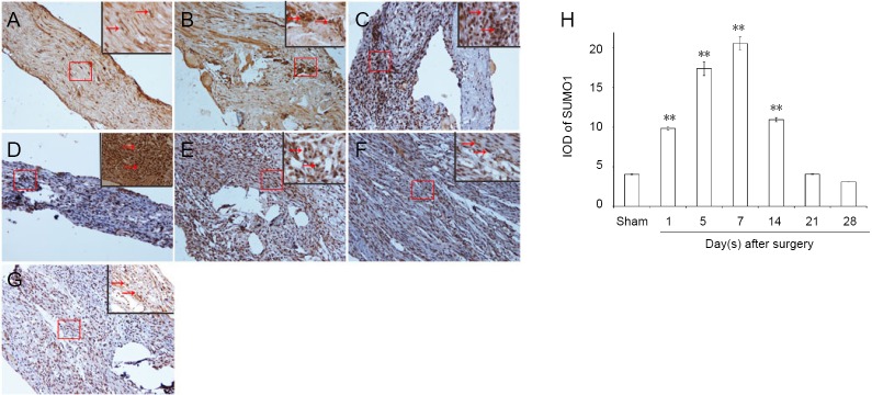
Figure 2.
Immunohistochemical staining for SUMO1 in sciatic nerves.
(A) SUMO1 immunopositive staining (arrows) in sciatic nerve of the sham group (1 cm sciatic nerves around the sciatic notch); (B–G) SUMO1 immunopositive staining (arrows) in the sciatic nerve (containing both 5 mm proximal and distal stumps at the injury site) at 1, 5, 7, 14, 21, and 28 days after sciatic nerve injury in the experimental group, respectively. SUMO1 immunopositive staining was observed in A–G, especially in the sciatic nerve suture point (the boxed areas) of Figure B–E. Using optical microscope, original magnification, 20×; insets are higher magnification of the boxed areas, original magnification, 100×. (H) Quantitative analysis of SUMO1 immunopositivity in each group. **P < 0.01, vs. sham group (mean ± SD, n = 4, one-way analysis of variance followed by Tukey’s honestly significant difference post hoc test). IOD: Integrated optical density; SUMO: small ubiquitin-like modifier.

