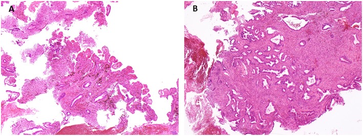
Figure 2.
Vaginal biopsies. The tissue transmitted for pathological analysis corresponds to endometrium, with numerous glands slightly irregular in size and shape lined by cylindrical cells without nuclear atypia, and densely cellular stroma with signs of ancient bleeding, rare mitotic figures, and no cytologic atypia. (A) Hemalun Eosin, magnification x4. (B) Hemalun Eosin, magnification x 10.

