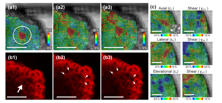
Fig. 3.
(a1-a3) Von Mises strain analysis and (b1-b3) corresponding fluorescence images of the further indentation sequence. When a large compression was added, cell displacement is observed as the balloon-like deflation and inflation from (b2) to (b3). The strain analysis detected the displacement at an early step of indentation in (a1). (c) Analysis of axial (x’), lateral (y’), and elevational (z) and shear (x’y’, zx’, and y’z) strain components. The direction of the indentation was chosen as the x’ axis. Scale bars = 50 µm. See also Visualization 1 (440.6KB, mp4) and Visualization 2 (236.6KB, mp4) .

