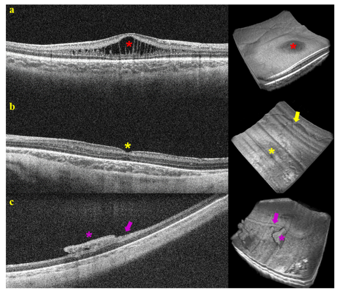
Fig. 7.
Selected B-scans and volume renders of the retina from research HH-SSOCT imaging of non-sedated infants. a) In a premature-born infant imaged in the outpatient clinic one week after estimated date for term birth, CME elevates the central macula (red star). b) In a premature infant imaged in the intensive care nursery weeks before estimated date for term birth, the fovea has a normal depression without edema (yellow star), but there are large superficial vessels on the surface of the retina (yellow arrow). c) Peripheral images from the same infant as b) reveal preretinal neovascular elevations (purple star) and a ridge at the vascular/avascular junction (purple arrow). All images were acquired at 950 A-scans/B-scan and 128 averaged B-scans/volume. Each B-scan was averaged twice.

