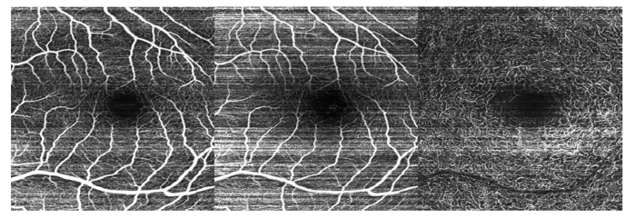
Fig. 10.
Imaging of vascular plexuses with HH-OCTA in a child undergoing EUA due to a family history of familial exudative vitreoretinopathy. Left: Projection of all vascular layers in the outer retina. Center: Projection of the SVC. Right: projection of the DVC. Images show relatively normal vascular patterns in each projection and were taken with 500 A-scans/B-scan, 4 repeated B-scans, and 500 lateral locations sampled per volume.

