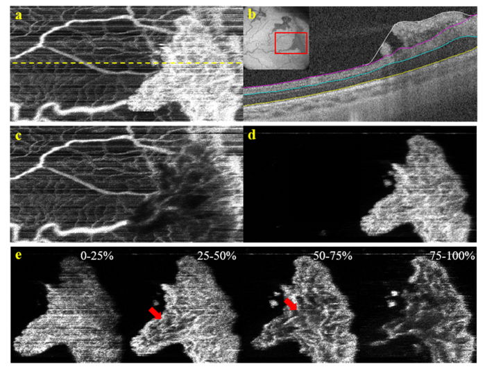
Fig. 12.
Vascular structure of a preretinal neovascular elevation in a 41 week PMA infant who was treated for ROP. Infant was imaged without sedation in the ICN. a) OCTA image of all retinal vasculature. Yellow line denotes the location of the B-scan shown in (b). b) Selected B-scan with manually corrected segmentations. White – surface of the retina including the neo-vascular plaque. Purple – surface of the retina excluding the neo-vascular plaque. Teal – IPL. Yellow – RPE c) OCTA of the vasculature excluding the neo-vascular plaque (purple to yellow). d) OCTA of the neo-vascular plaque (white to purple). e) The neo-vascular plaque divided into slices that are 25% of the total thickness of the plaque. The red arrow denotes the location of the “trunk” within the neovascular plaque.

