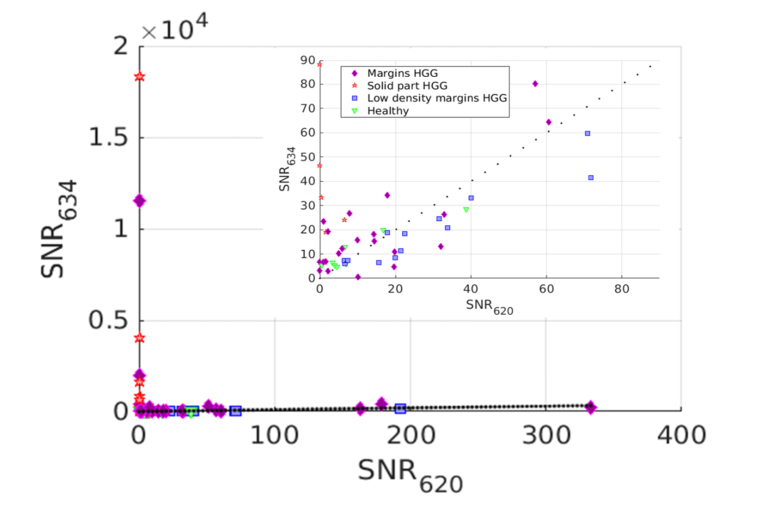
Fig. 5.
SNR634 versus SNR620 for HGG, with a zoom. Markers indicate anatomo-histopathological classification: solid part of HGG (red stars), HGG margins (orange diamonds), HGG margins with low density of tumor cells (blue squares) and healthy tissues (green triangles). Dotted line shows the equality of both SNR contributions.

