Fig. 9.
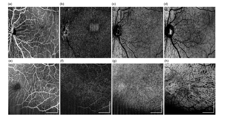
OCTA imaging results can be obtained from premature infants. Results were obtained from an infant at gestational age of 40 weeks, with a FOV of 7.0 x 7.0mm: (a-d) OCTA maximum intensity projection en face images produced from four layers, superficial retina, deep retina, choriocapillaris, and deep choroid, respectively. (e-h) higher magnification OCTA en face images were obtained from an infant at gestational age of 28 weeks, with a FOV of 3.6 x 3.6mm, which are presented in the same order as the row above. All scale bars: 1 mm.

