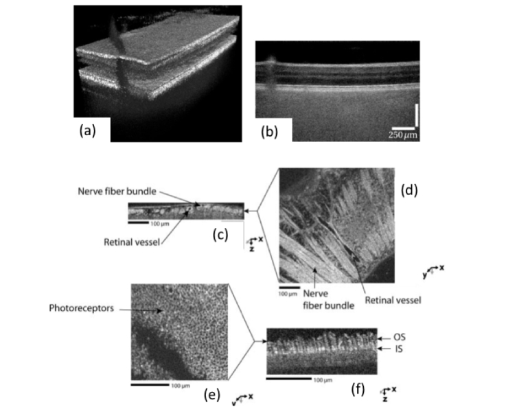
Fig. 8.
Imaging examples for FF OCT: SS FF OCT in-vivo results of human retina showing (a) a rendered 3D volume and (b) a corresponding tomogram (10 times averaged) exhibiting good contrast for the inner retinal layers and loss of contrast for the choroidal structures (reproduced from [88] with permission of The Optical Society); (c-f) TD FF OCT results of ex-vivo retinal tissue (Republished with permission of the Association for Research in Vision and Ophthalmology from [5]; permission conveyed through Copyright Clearance Center, Inc.)

