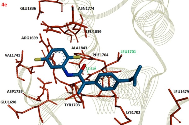
Figure 3.

Docking complex of 4e against BRCA1.
Notes: The ligand structure is shown in Berghaus blue while the functional groups such as oxygen, nitrogen and chlorine are displayed in red, blue and yellow colors, respectively. The active site residues are depicted in dark red and labeled with black. The amino acid which participates in hydrogen bonding is highlighted in green along with bonding distance in angstrom (Å). The receptor protein is displayed in line format in light grey.
