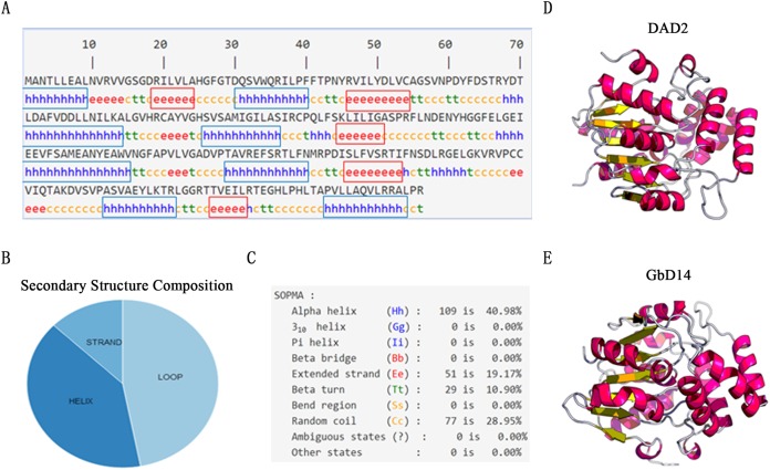
Figure 2. Advanced structure prediction of GbD14.
(A) Distribution of GbD14 secondary structure. “h” stands for alpha helix and is marked with a blue box. “e” stands for extended strand and is marked with a red square. (B) Composition of GbD14 secondary structure (strand, loop, helix). (C) Proportion of GbD14 secondary structure. The three-dimension structures of DAD2 (D) and GbD14 (E).

