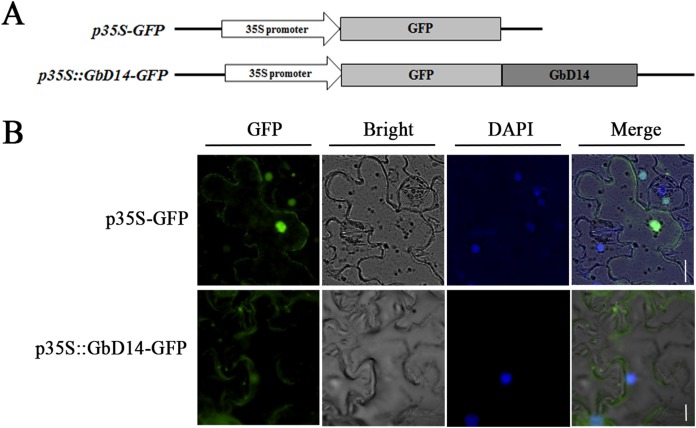
Figure 4. Subcellular localization of GbD14 in tobacco.
(A) Schematic diagram of p35S-GFP::GbD14. Drawing is not to scale. (B) Subcellular localization of GbD14 in a tobacco epidermal cell. DAPI labels the location of nucleus. Transient GbD14-GFP fusion proteins was visualized 48 h after agroinfiltration. Bars, 20 μm.

