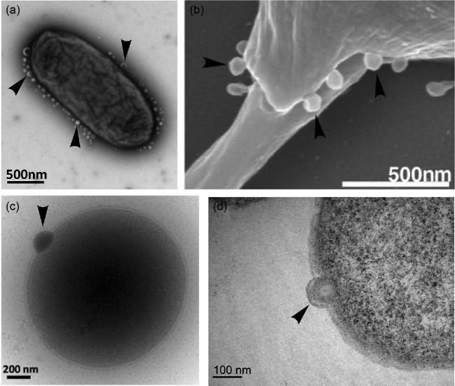
Figure 1.
Biogenesis of extracellular vesicles in the three domains of life. Vesicle budding indicated with arrows. (a) TEM showing hypervesiculation in the bacterium S. typhimurium. Image kindly provided by Mario F. Feldman (University of Alberta, Canada). (b) SEM showing microvesicles budding from the eukaryote Leishmania donovani. Image reprinted from Silverman et al. (2008). (c) Cryo-TEM of vesicle budding from the archaeon T. kodakaerensis. The protrusion of the S layer can also be observed clearly. (d) TEM of ultrathin cell sections of vesicle budding from T. kodakaerensis. Figures (c) and (d) provided by the authors.

