Fig. 3.

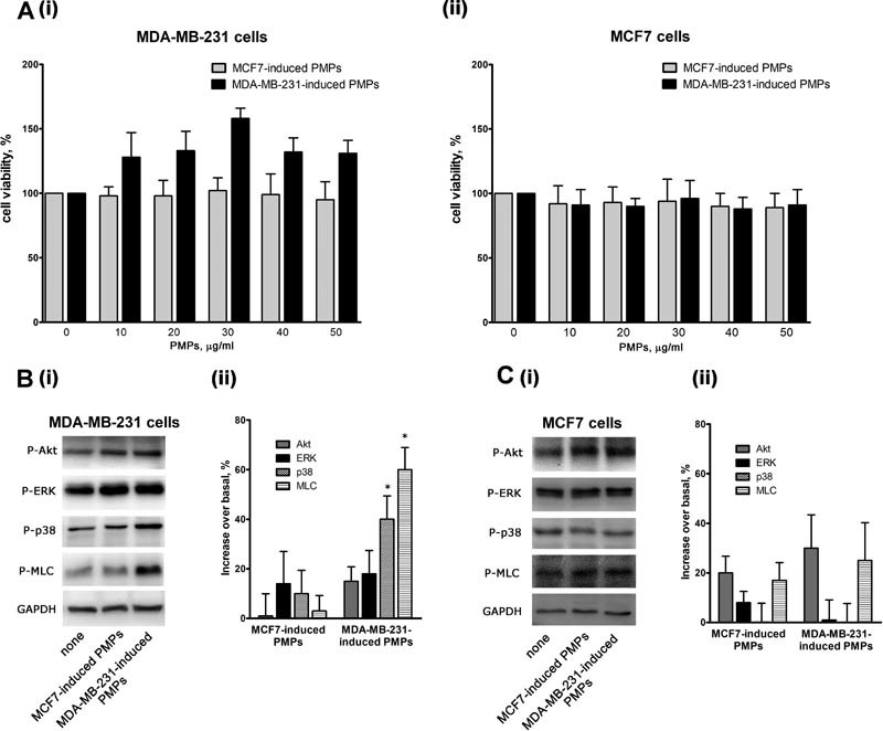
Platelet-derived microparticles (PMPs)-induced activation of MDA-MB-231 cells. ( A ) Viability of MDA-MB-231 (i) or MCF7 (ii) cells incubated with the indicated amounts of PMPs for 24 hours was assessed by a colorimetric MTT assay. Results are reported as the mean ± SEM of three different experiments. ( B and C ) Phosphorylation of selected signaling proteins in MDA-MB-231(panel B) or MCF7 (panel C) cells incubated with MCF7- or MDA-MB-231-induced PMPs for 18 hours, as indicated on the bottom. Representative immunoblot with specific anti-phosphoprotein antibodies directed against the protein indicated on the right is reported in (i), where GAPDH staining is for equal loading control. Quantification of the results by densitometric scanning is reported in (ii), as % of phosphorylation increase over the level of untreated cells. Results are the mean ± SEM of three different experiments. * p < 0.05.
