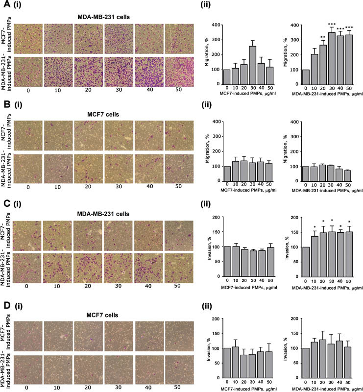
Fig. 4.

Analysis of cell migration and invasion. Effect of MCF7- or MDA-MB-231-induced PMPs (as indicated on the left) on migration (panels A and B) and invasiveness (panels C and D) of MDA-MB-231 cells (panels A and C) or MCF7 cells (panels B and D), as indicated. Cancer cells were treated with increasing amounts of the two different types of PMPs preparations (0–50 μg/mL, as indicated on the bottom) and then transferred inside cell culture inserts. For the invasion assays (panels C and D), the upper side of the insert was coated with 0.1 mL of Matrigel (50 µg/mL). Incubation was prolonged for 18 hours and the cells that moved through the porous membrane were stained and counted. In all the panels, representative images are reported in (i), while quantification of the results is shown in (ii) as mean ± SEM of three experiments. Statistical significance of the difference was calculated between treated and untreated cells (sample 0). * p < 0.05; ** p < 0.01; *** p < 0.001.
