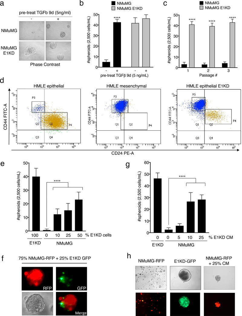
Figure 1. TGFβ/hnRNP E1 induces BCSC Self-Renewal through a Secreted Factor.
(a) Phase contrast images and (b) quantification of mammosphere formation by NMuMG and NMuMG shE1 (E1KD) cells with or without 9-day TGFβ pre-treatment (error bars represent mean +/− SD; n=5; ****p <0.0001, unpaired Student’s t-test). All mammospheres were counted at a minimum diameter of 100μm. (c) Quantification of sequentially passaged mammospheres from NMuMG and E1KD cells (error bars represent mean +/− SD; n=5; ****p <0.0001, unpaired Student’s t-test). (d) FACS analysis of CD44/CD24 surface marker expression in HMLE epithelial, mesenchymal, and epithelial E1KD populations (HMLE epi P3: 3.4%, P4: 92.7%; HMLE mes P3: 98.0% P4: 0.7%; HMLE epi E1KD P3: 80.8%, P4: 12.5%) (e) Mammosphere quantification and (f) representative images from co-cultured NMuMG-RFP and E1KD-GFP cells as well as (g & h) NMuMG cells supplied with increasing amounts of E1KD conditioned media (error bars represent mean +/− SD; n=10; ****p <0.0001, One-way Anova.

