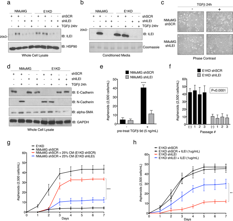
Figure 2. The Secreted Cytokine ILEI is Necessary for TGFβ/hnRNP E1-Mediated EMT and BCSC formation.
Immunoblot analysis of either (a) whole cell lysates or (b) conditioned media from NMuMG and E1KD cells stably transduced with either scramble control shRNA (shSCR) or ILEI knock-down shRNA (shILEI) in the presence or absence of TGFβ (5ng/ml) for 24 hours. Coomassie staining was used as a loading control for conditioned media. (c) Phase contrast images of NMuMG shSCR and NMuMG shILEI cells in the presence and absence of TGFβ stimulation for 24 hours. (d) Immunoblot analysis of EMT markers in whole cell lysates derived from NMuMG/E1KD shSCR and NMuMG/E1KD shILEI cells in the presence and absence of TGFβ stimulation for 24 hours. (e) Quantification of mammosphere formation by TGFβ pre-treatment (5ng/mL) in NMuMG cells stably transduced with either scramble control shRNA (shSCR) or ILEI knock-down shRNA (shILEI) (error bars represent mean +/− SD; n=5; ****p<0.0001, unpaired Student’s t-test). (f) Quantification of mammosphere formation and self-renewal in E1KD cells stably transduced with either scramble control shRNA or ILEI knock-down through several passages (error bars represent mean +/− SD; n=5, p <0.0001 for all passages, paired Student’s t-tests between correlative passage numbers). (g) Mammosphere formation by NMuMG shSCR cells supplemented with conditioned media derived from either E1KD shSCR or E1KD shILEI cells (25% total volume) with E1KD mammosphere growth used as a positive control (error bars represent mean +/− SD, n=5; ***p=0.0002, 2-way ANOVA). (h) Quantification of mammosphere formation in E1KD shSCR/shILEI cells in the presence and absence of purified recombinant ILEI (error bars represent mean +/− SD; n=5; **p=0.0013, 2-way ANOVA).

