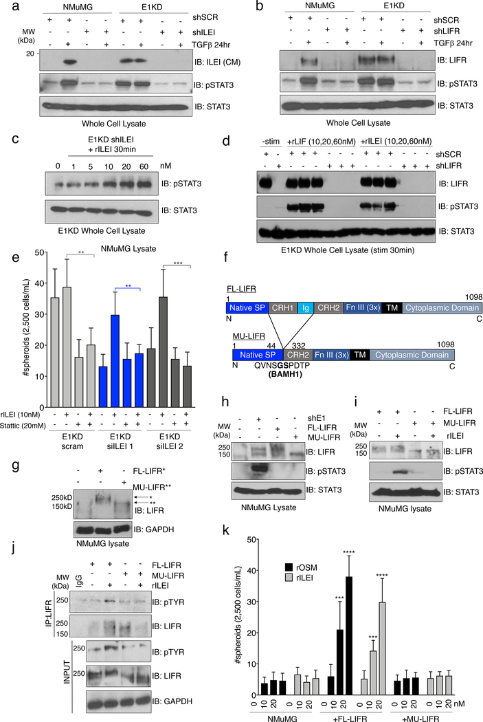
Figure 5. ILEI Induces STAT3 Signaling through Activation of LIFR.
(a) Immunoblot analysis of basal pSTAT3 levels in NMuMG and E1KD cells stably transduced with control shSCR shRNA, as well as shRNA targeting either ILEI or (b) LIFR. Cells were stimulated with TGFβ (5ng/mL) for 24 hours where indicated. (c) Stimulation of E1KD shILEI cells with the indicated concentration of recombinant ILEI for 30 minutes. Lysates were probed for phosphorylated STAT3 protein. (d) E1KD cells or E1KD shLIFR cells treated with increasing concentration of rLIF and rILEI for 30 minutes. Lysates were probed for LIFR, pSTAT3, and total STAT3. (e) Mammosphere assay using E1KD cells treated with two siRNA molecules against ILEI and rescued with rILEI treatment at 10nM. The STAT3 inhibitor Stattic was added at 20μM where indicated (error bars represent mean +/− SD; n=5; **p<0.01, ***p<0.001, unpaired Student’s t-test). (f) Construct diagram of FL-LIFR and mutated LIFR (MU-LIFR) lacking the cytokine binding region. Regions include the native signal peptide, cytokine receptor homology domains 1 and 2, Ig-like domain, Fibronectin type III repeat, transmembrane and cytoplasmic domains. (g) Overexpression of FL-LIFR and MU-LIFR constructs in NMuMG cells. (h) Immunoblot analysis of basal STAT3 activation in NMuMG cells overexpressing either FL-LIFR or MU-LIFR compared to parental NMuMG and E1KD cells. (i) Immunoblot analysis of STAT3 activation in response to rILEI (20nM) for 30 minutes in either NMuMG FL-LIFR or MU-LIFR cells. (j) Immunoprecipitation of LIFR after rILEI stimulation (20nM) for 30 minutes in NMuMG cells overexpressing either FL-LIFR or MU-LIFR followed by immunoblot analysis. (k) Quantification of mammosphere formation in NMuMG FL/MU-LIFR cells in the presence or absence of rOSM/rILEI pre-treatment for 7 days (error bars represent mean +/− SD; n=5; ****p<0.0001, ***p<0.001, unpaired Student’s t-test).

