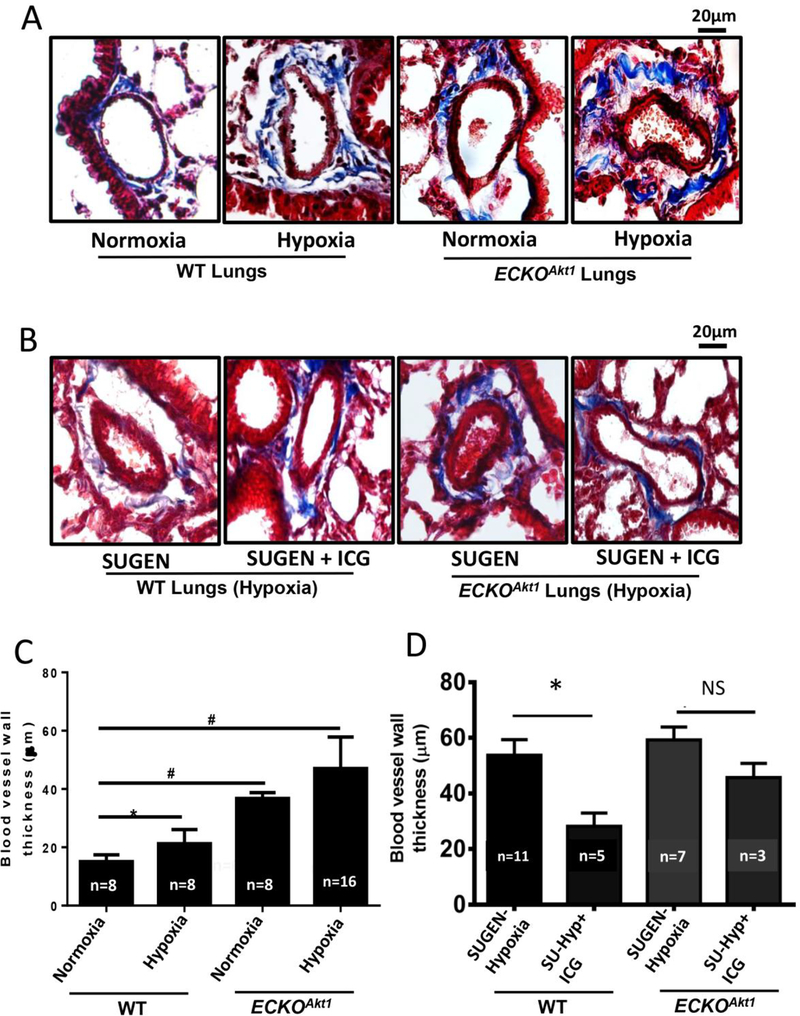
Figure 4. Endothelial loss of Akt1 exacerbates hypoxia-induced vascular remodeling in vivo through β-catenin.
(A) Representative images of Masson’s trichrome staining and (C) Bar graph indicating the blood vessel thickness of lung sections of WT and ECKOAkt1 mice subjected to either normoxia or 10% hypoxia for 3 weeks. (B) Representative images of Masson’s trichrome staining and (D) Bar graph indicating the blood vessel thickness of lung sections of WT and ECKOAkt1 mice injected with SUGEN and subjected to hypoxia for 3 weeks. The treatment group received ICG001, a β-catenin inhibitor. Vessel thickness was measured using NIH Image J software. One-way ANOVA was performed and data are represented as mean ± SD. *p<0.05; #p<0.01.

