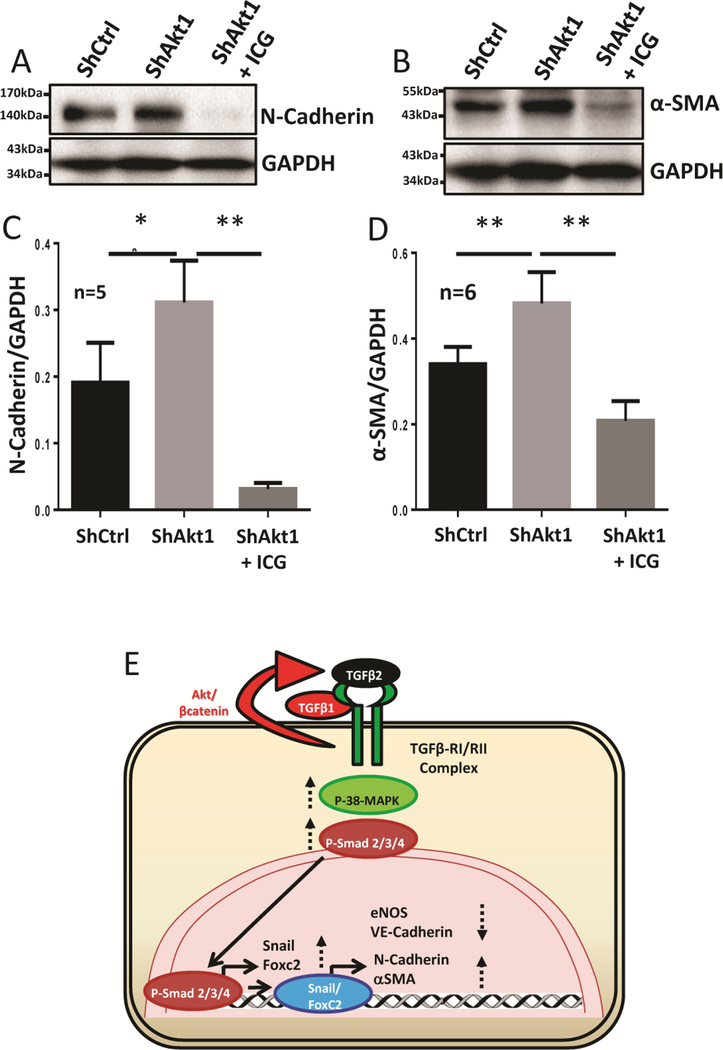
Figure 6. EndMT in ShAkt1 HMECs is blunted by treatment with β-catenin inhibitor ICG-001.
(A-B) Representative Western blot images of untreated ShControl and ShAkt1 HMECs, as well as ShAkt1 HMECs, treated with ICG-001 indicating the changes in the expression of mesenchymal markers N-cadherin and αSMA. (C-D) Bar graph showing the changes in the expression of mesenchymal markers N-cadherin and αSMA in untreated ShControl and ShAkt1 HMECs, as well as ShAkt1 HMECs, treated with ICG-001. (E) Schematic diagram showing the working hypothesis on the molecular mechanisms by which endothelial loss of Akt1 contributes to EndMT. One-way ANOVA was performed and data are represented as mean ± SD. *p<0.05; **p<0.01.

