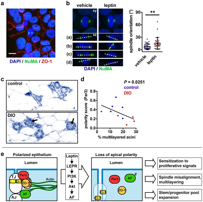
Fig. 6.
Leptin leads to mitotic spindle misalignment. a Detection of ZO-1 and NuMA by immunofluorescence in polarized monolayers of S1 cells. Scale bar, 10 µm. b Mitotic spindle orientation in S1 cell monolayers treated with vehicle or leptin, as measured by confocal microscopy. Images on top represent maximal intensity projections whereas bottom images (a-d) are representative orthogonal views of the dual NuMA-DAPI staining. Angles between the two spindle poles (dashed lines) and the horizontal are quantified in the graph. **, P < 0.01 (unpaired t-test, n = 30 cells from two independent experiments). c Histology of mammary glands from mice with diet-induced obesity (DIO) and mice fed a control diet. Arrows indicate multilayered acini, defined as having ≥ 3 cell layers. d Correlation between apical polarization (Par3 score) and multilayering in the mammary epithelium from control and DIO mice. The exact P value (two-tailed Spearman test) was computed. The graphs represent mean ± SEM (B-E) or mean ± SD (G). e Schematic summarizing the effect of leptin on epithelial polarity and the functional consequences of polarity loss in the premalignant context.

