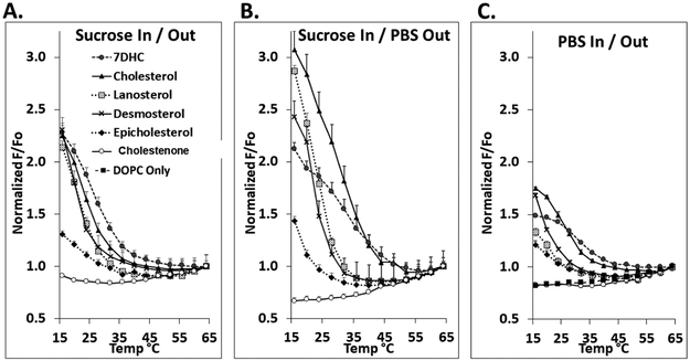
Figure 3.
Domain melting curves in symmetric vesicles assayed by FRET. The fraction of DPH fluorescence unquenched by rhodamine lipid (F/Fo) versus temperature shown after normalization to F/Fo at high temperature (64°C). Samples contained LUV with 100 μM lipid composed of 1:1 mol:mol DOPC:SM with 25 mol% sterol. Samples contained 0.01 μM DPH, and ‘F samples’ also contained 2 mol% rhodamine-DOPE. In panels A. and B., vesicles were formed with entrapped sucrose and dispersed in either sucrose (A.) or PBS (B.). In C., vesicles were formed with entrapped PBS and dispersed in PBS. Mean and standard deviation from three separate experiments are shown. Symbols: Filled circles, 7DHC; triangles, cholesterol; shaded squares, lanosterol; crosses, desmosterol; diamonds, epicholesterol; open circles; 4-cholesten-3-one; filled squares, no sterol.

