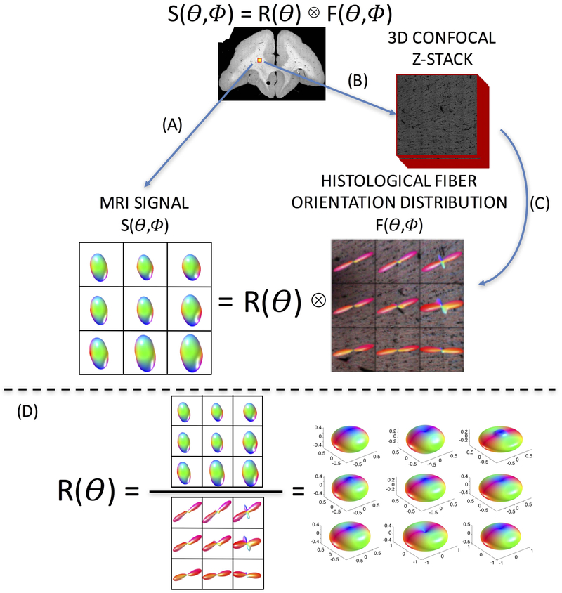
Figure 1.
Overview of study methodology. Spherical deconvolution models the diffusion signal, S, as the convolution of the fiber response function, R, with the fiber orientation distribution, F. The brain is scanned (A) resulting in an MRI signal. Next, 3D confocal microscopy is acquired (B), followed by image processing (C), resulting in the ground truth histological FOD. Estimating the voxel-wise response function (D) is done through deconvolution of the signal with the corresponding FOD.

