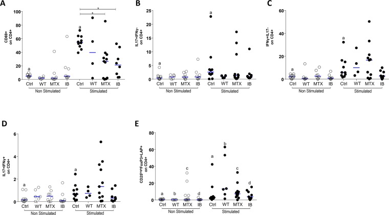
Figure 4.
Flow cytometric analysis of cell activation markers and intra-cellular cytokine expression on CD4+ T-lymphocytes (TL): The PBMCs were obtained from healthy subjects (Ctrl), psoriasis patients without therapy (WT), patients treated with methotrexate (MTX), and patients treated with immunobiologicals (IB). PBMC, incubated for 48 h in the presence of medium (unstimulated) or with anti-CD3 and anti-CD28 mAbs (stimulated). After recovered, PBMC were incubate with appropriate mAbs and isotype controls. Panel A: CD69+ on CD4+ TL (aWilcoxon; p = 0.05 and *Kruskal-Wallis p < 0,05), panel B: IL-17+IFN-γ− on CD4+ LT (aWilcoxon p > 0.05), panel C: IL-17− IFN-γ+ on CD4 + LT (aWilcoxon p < 0.05), panel D: IL-17+IFN-γ+ on CD4 + LT (aWilcoxon p > 0.05) and panel E: CD25+ FoxP3+LAP+ on CD4+ LT (aWilcoxon; p 0.05 and *Kruskal-Wallis p < 0,05). The results are expressed in percentage on the CD4+ TL. A and *indicates statistical significance.

