Fig. 6.
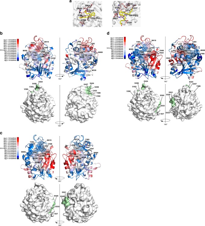
In silico analyisis of the allosteric communication induced by perturbation of the reactive cysteine residues of hsEH CTD. a Binding poses of 15d-PGJ2 at the predicted C423 (left) and C522 (right) binding sites. The ligand is depicted in yellow, while the interacting residues are depicted in brown. b–d AlloSigMA analysis of the perturbation of binding sites (C423, C522, C423 and C522 respectively). The top panels show the cartoon model of hsEH CTD, coloured according to the predicted Δgi (blue: destabilization—Δgi ≤ −0.2 kcal/mol; red: stabilisation—Δgi ≥ 0.2 kcal/mol). The bottom panels depict the surface representation, with the perturbed binding sites in green. The full Δgi profiles are reported in Supplementary Fig. 8

