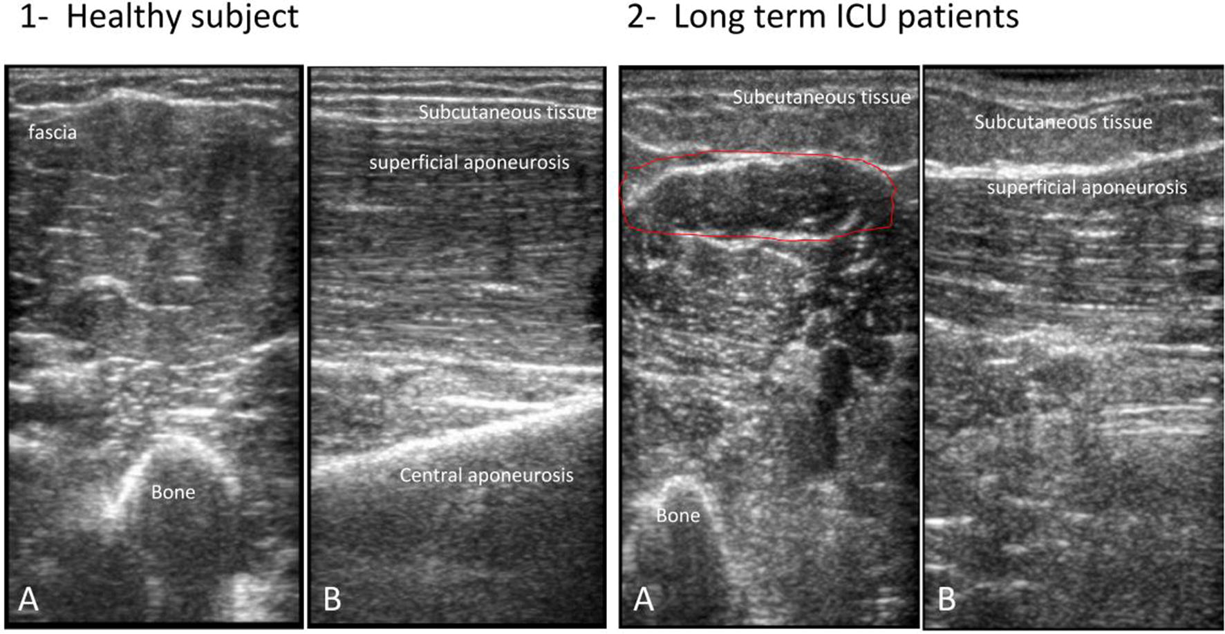
Fig. 1.

Ultrasound distinctive appearance of muscle tissue. The figure shows a transverse (a) and longitudinal (b) ultrasound scan of elbow flexor (bicep brachialis) in healthy (1) and long-term ICU (2) subjects. In the axial image, muscle consists of primarily hyper-echogenic areas scattered with small bright curved echoes of superficially random orientations. In the sagittal plane, these bright echoes are seen to be the fibrous tissue that surrounds muscle fibers and fascicles and which organize into recognizable striations. In bipennate or multi-pennate muscles, a central aponeurosis can be identified as an area of thickened fibrous tissue that when followed distally becomes the tendon. Bone is highly echogenic with a deep shadow beneath the bright hard edge. Subcutaneous fat is typically of similar echogenicity to muscle and is interposed with brighter, poorly organized strips of connective tissue. Near the myotendinous junction, the myofascial fibrils merge, resulting in increased echogenicity and higher anisotropy. In the healthy tissue, the hyper-echogenic muscle is interspersed with bright fibro adipose tissue and the bone reflection is bright and sharply defined; in the long-term ICU patient, the muscle tissue appears as non-homogenous and reduced in its mass
