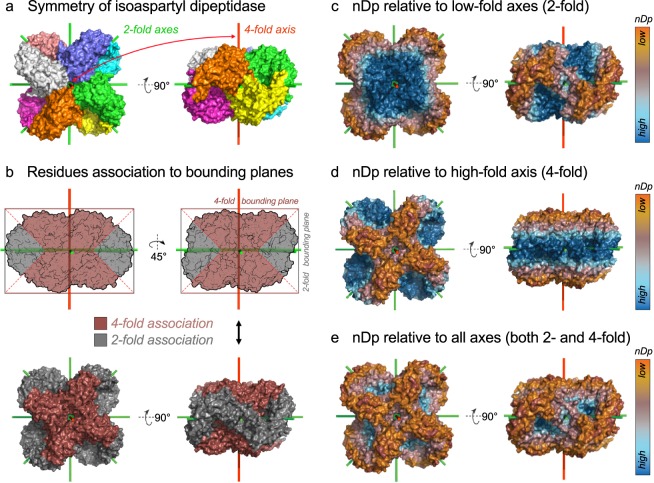
Fig. 1.
Principle of calculation of different versions of the normal distance to the closest bounding plane (nDp) visualized on the dihedral structure of isoaspartyl dipeptidase. (a) Coloration of the biological assembly of isoaspartyl dipeptidase by subunits (PDB accession 1POK35). Symmetry axes appear in green (2-fold axes) and red (4-fold axis). (b) Residues are assigned to their closest bounding plane. For this D4 complex, bounding planes originate from either 2- or 4-fold axes (grey and brown, respectively). (c) Visualization of the nDp-2-fold. (d) Visualization of the nDp-n-fold, where n = 4 in the case of this D4 complex. (e) Visualization of the nDp, which is relative to all bounding planes of the assembly independently of axes folds.

