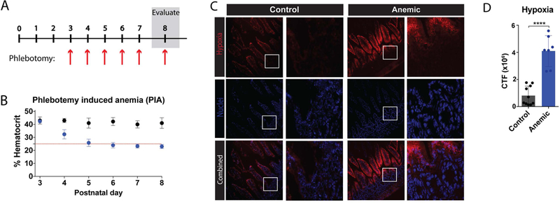
Figure 2. Phlebotomy induced anemia (PIA) results in increased hypoxia within the intestinal mucosa.

(A) Schematic of protocol for induction of PIA. (B) Percent hematocrit of neonates with (blue) or without (black) phlebotomy on days 3–8 after birth (P3-P8) as indicated. Red line indicates threshold for anemia (25%). (C) Fluorescence microscopy imaging of intestines isolated from control or anemic pups at P8, following administration of Hypoxyprobe (Pimonidazole HCl), which forms adducts in hypoxic tissue. Frozen tissue was stained for tissue hypoxia (pimonidazole) (red) and nuclei (DAPI) (blue). White boxes indicate enlarged regions shown to the right of each 20x field. (D) Quantification of fluorescent hypoxia in 2C. CTF = corrected total fluorescence. Data are presented as mean ± SEM, ****P < 0.0001.
